The spinal meninges and associated structures Adipose tissue space Epidural space Dura mater Dorsal root ganglion Spinal cord Dorsal root Arachnoid mater Ventral root Pia mater. IAC Lab AP I WEDS T130am-140pm Hi Vanessa v Sign Out Help Mastering AP Course Home My.
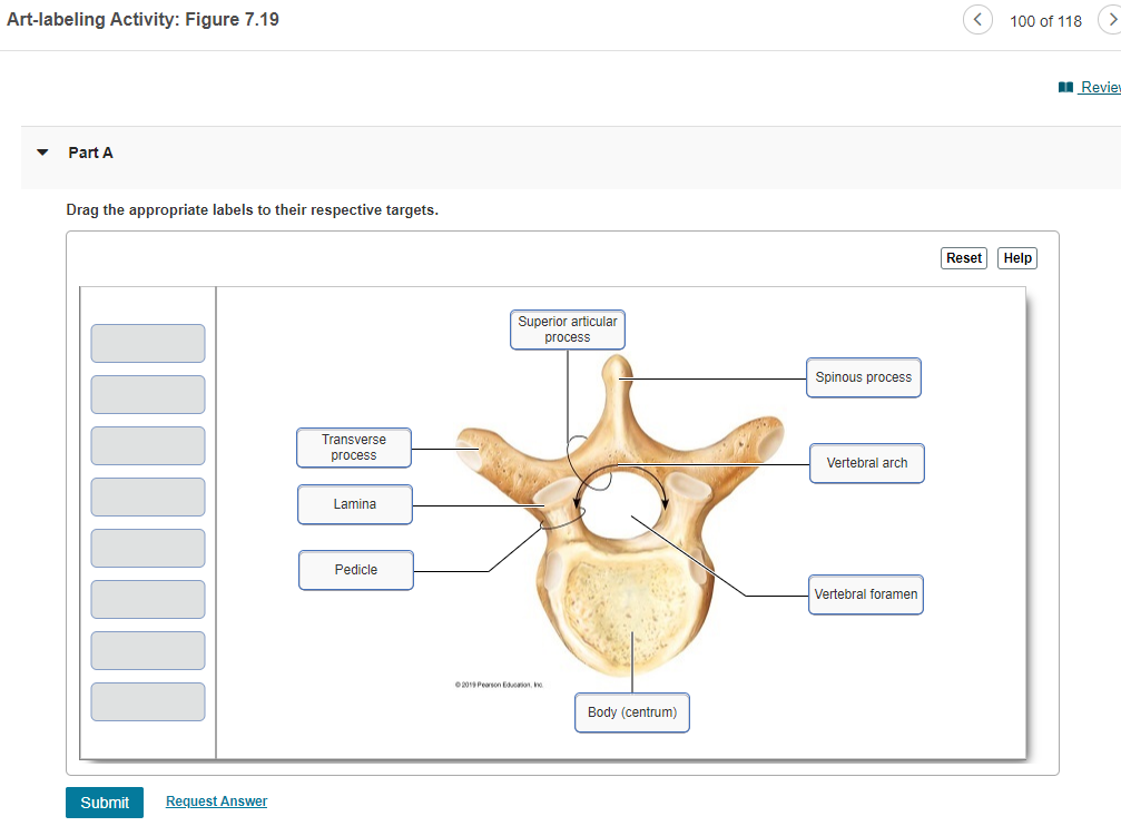
Solved Art Labeling Activity Figure 7 19 100 Of 118 Revie Chegg Com
Start studying BIO2341-181 Gross Anatomy of the Heart Art-Labeling Activity.
. The Cellular Level Of Organization Review Guide. External anatomy of the heart anterior surface Art-labeling Activity. Figure 137 Label the Spinal Nervesfront 2 back 2 front 3 back 3 front 4 back 4.
Learn vocabulary terms and more with flashcards games and other study tools. It runs along the vertical axis of the body. The Spinal Cord and Spinal Meninges.
Homework for A P. BIO 333 Chapter 15 WS. THIS SET IS OFTEN IN FOLDERS WITH.
Lower trunk formed by the joining of roots C8 and T1. Like the dorsal cavity the ventral cavity has two subdivisions. Anatomy and Physiology 1.
The structure indicated by label e is part of which of the following. The manubrium the body. The Spinal Cord and Spinal Meninges.
The appendicular region includes the limbs which are also. An 11-page dissection guide that covers the internal and external anatomy of the sheep heart. Structure of the epidermis part a drag the appropriate labels to their respective targets.
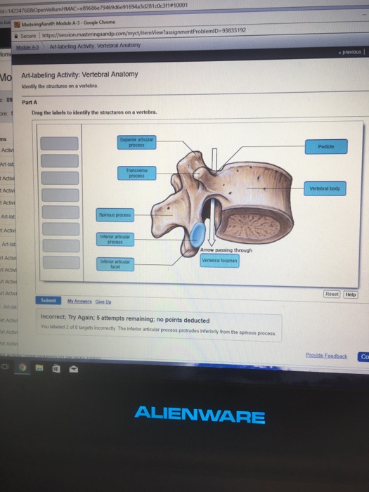
Nail Structure - Art Labeling Activity Vertebral Anatomy Jpg M No Subject Stellarvore91 Gm X G Anatomy Abdominal Quadrants X Course Home X Smart Pearson Course Hero - Reset help nail root of nai plate nail body of nail plate prodmal nail fold hyponychium lateral nail foid eponychium nail bed lunula nail matri medial nail fold free adge of. Instructors may assign this figure as an Art Labeling Activity using Mastering APTM Regional Anatomy The body is divided into two main regions the axial and appendicular regions. Bones of the Adult Skull Anterior View Part 2.
Art Labeling Activity. Brain Cranium and Meninges Close-up View of Cranial Meninges Part A Drag the labels to the appropriate location in the figure. Figure 1311b Label the nerves on.
English French German Latin Spanish View all. So make sure you learn from the feedback. The seven bones of the neck are called vertebrae.
Exercise 19 Review Sheet. The spinal cord and spinal meninges. May 07 2022.
View art labeling activity - anatomy of a thoracic vertebrajpg from EGN MISC at Miami Dade College Miami. Curves and Regions of the Vertebral Column Sacral curve 7 vertebrae Sacral region Lumbar region Coccygeal region Thoracio region 12 vertebrae To VIO Cervical region Thoracic curve 13 Il 1 Cervical curve 5 vertebrae Lumbar curve. Basic Anatomy Of The Skin.
The Organ Systems Part 2 Label the diagram to identify the organ systems 35 terms. AP II LAB Heart Anatomy terms. Like the dorsal cavity the ventral cavity has two subdivisions.
The meninges are the protective coverings which enclose the brain and spinal cord. Match each anatomical term in the key to the. The anterior horns are wider than the posterior horns.
Of the three layers of the meninges the dura mater the. See Diagram Art-labeling Activity. The spinal vertebral cavity encases the vertebral column and spinal cord.
The axial region includes the head neck and trunk. The Spinal Cord and Spinal Nerves Anatomy of the Spinal Cord 1. Instructors may assign this figure as an art labeling activity using mastering aptm regional anatomy the body is divided into two main regions the axial and appendicular regions.
The three major sensory tracts involve chains of neurons First-order neuron Delivers sensations to the CNS The cell body is in the dorsal or cranial root ganglion Second-order neuron An interneuron with the cell body in the spinal cord or brain Third-order neuron Transmits information from the thalamus to the PDF The. Reset Hel Supraspinous ligament Primary Vertebral Ligaments Intervertebral disc. Figure 137 Label the Spinal Nerves See Diagram 5 Art-labeling Activity.
A set of venous chain that are located between 2 layers of dura mater and drain the cerebral veins of the brain. Start studying Art-Labeling Activity. Start studying Art-Labeling Activity.
Heart Anatomy Practice Quiz. Lines and Cues Narrator 25 terms. The vertebrae articulate with the corresponding ribs.
Vertebral Into the Woods. The commonly referred to as the breastbone is a flat bone formed by the fusion of three bones. The epithelium is a type of body tissue that forms the covering on all internal and external surfaces of your body lines body cavities and hollow organs and is the major tissue in glands.
Anatomy and Physiology questions and answers. Primary vertebral ligaments and structural features of the intervertebral disc Part A Drag the labels onto the diagram to identify the primary vertebral ligaments and structural features of the intervertebral disc. The appendicular region includes the.
Soon after their origin these 5 nerve roots unite to form three trunks.

Art Labeling Activity Vertebral Anatomy Jpg M No Subject Stellarvore91 Gm X G Anatomy Abdominal Quadrants X Course Home X Smart Pearson Course Hero

Vertebral Anatomy A Lateral And Slightly Inferior View Of A Vertebra Diagram Quizlet
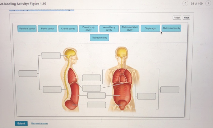
Solved Art Labeling Activity Figure 1 10 93 Of 109 Reset Chegg Com

Vertebral Anatomy Anatomy Bones Medical Anatomy Human Anatomy And Physiology
Solved E89686e79469d6e91694a3d281c0c3r1 10001 D 14234 Chegg Com
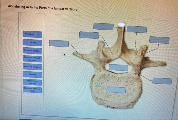
Solved Art Labeling Activity Parts Of A Lumbar Vertebra Chegg Com

Thoracic Vertebrae Human Anatomy And Physiology Body Anatomy
Solved Skeletal System Hw Art Labeling Activity Vertebral Chegg Com
0 comments
Post a Comment